Biological complexity and the challenges associated with its elucidation
Biological organisms are complex systems composed of a variety of cell and tissue types.1 To elucidate the physiological and pathophysiological processes that occur in biological systems, research studies should take into consideration their complexity. However, many traditional experimental paradigms either do not allow the proper evaluation of this biological complexity or do not enable a fast and scalable assessment. Molecularly barcoded (magnetic) microparticles (beads) are one of the avenues to advance research on biological complexity.
Molecularly barcoded beads and their applications
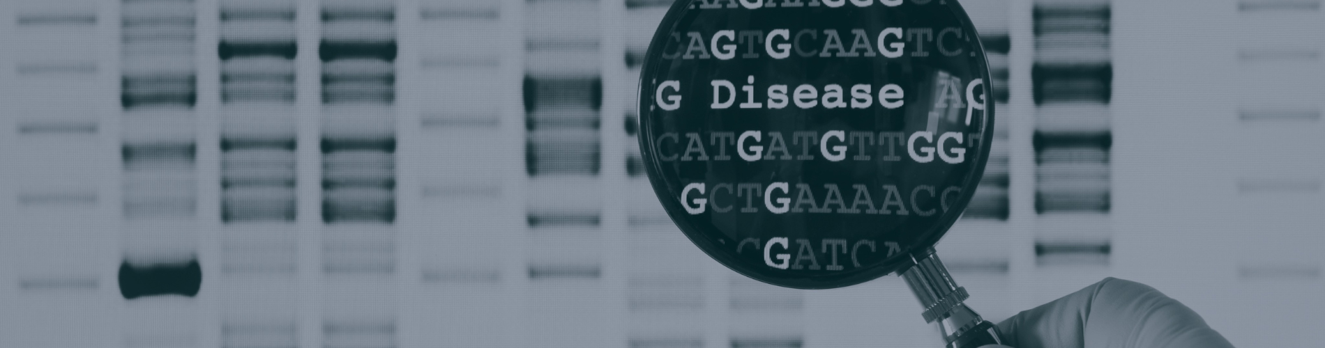
Molecularly barcoded beads are tagged with unique barcodes (molecules) that enable single-cell evaluation. The number of codes varies depending on the used assay and may include fluorescent, chemical, electronic, or graphic tagging. Molecularly barcoded beads facilitate the identification of reads originating from individual cells. In the context of microfluidic systems, they enable flexibility, sensitivity, and high-throughput analysis. Therefore, barcoded beads have found applications in multiplex biological assays in the fields of molecular diagnostics, biomarker development, analysis of tumor heterogeneity, pharmacogenomics, understanding of tissue complexity, and gene mutational analysis.
Oligonucleotide-barcoded beads
Oligonucleotides are one of the types of molecular bead barcodes. The design of oligonucleotide-barcoded beads used with the Drop-Seq method has been described by Macosco et al. (2015).2 The authors synthesized oligonucleotide primers on the beads in the 5′ to 3′ direction, which yields free 3′ ends available for enzymatic priming. The oligonucleotide tags consist of four parts: a constant sequence, a “cell barcode”, a unique molecular identifier, and a capturing and priming oligo-dT sequence. The constant sequence serves as a priming site for downstream PCR and sequencing. The “cell barcode” is identical on the surface of each bead across all primers but differs from the cell barcodes present on other beads. The unique molecular identifier differs among primers and identifies PCR duplicates.3 Finally, the oligo-dT sequence is responsible for capturing polyadenylated mRNAs and priming reverse transcription.2
Oligonucleotide-barcoded beads in the context of the Drop-Seq technology
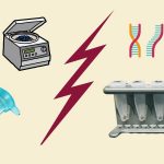
When molecularly barcoded beads are applied in the context of sequencing, they enable tracing the sequence back to the individual cells. The Drop-Seq technology is a microfluidic-based experimental system, in which beads tagged with oligonucleotides facilitate the retrieval of single-cell transcriptomes.4 Using a microfluidic device, single cells are encapsulated together with barcoded beads and lysis buffer into aqueous droplets. Following encapsulation, the cells are lysed, and mRNA is released and hybridized to the oligonucleotide tags of the beads. Subsequently, the droplets are pooled and broken, and the beads are released. The single-cell mRNAs captured on the isolated beads are then subjected to reverse transcription with template switching. cDNAs are generated and amplified, and sequencing adapters are added. Finally, the generated barcoded mRNA samples can be sequenced.2,4 More recently, methods for the identification and error-correction of molecular bead barcodes have been identified, including circularization of the RNA sequence.5
Overall, the Drop-Seq method, which utilizes oligonucleotide barcoded beads, not only analyzes mRNA transcripts from thousands of individual cells but also registers their cells of origin. Thus, the method enables high-throughput, single-cell transcriptomic analysis, which facilitates the elucidation of biological complexity.
Sources:
- Lobo, I. Biological complexity and integrative levels of organization. Nature Educ. 2008;1(1):141.
- Macosko EZ, Basu A, Satija R, Nemesh J, Shekhar K, Goldman M, Tirosh I, Bialas AR, Kamitaki N, Martersteck EM, Trombetta JJ, Weitz DA, Sanes JR, Shalek AK, Regev A, McCarroll SA. Highly parallel genome-wide expression profiling of individual cells using nanoliter d Cell. 2015;161(5):1202-1214.
- Kivioja T, Vähärautio A, Karlsson K, Bonke M, Enge M, Linnarsson S, Taipale J. Counting absolute numbers of molecules using unique molecular identifiers. Nat Methods. 2011 Nov 20;9(1):72-74.
- https://www.dolomite-bio.com/how-it-works/drop-seq/
- Tambe A, Pachter L. Barcode identification for single cell genomics. BMC Bioinformatics. 2019;20(1):32.