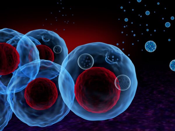
What are Exosomes?
Exosomes are extracellular vesicles which contain several cell components including proteins, DNA and RNA. Fusion of the plasma membrane with multivesicular bodies (MVBs) produces exosomes (Figure 1). These vesicles are mainly released by a cell to communicate or interact with neighboring cells by delivering information within DNA and several types of RNA, for example, microRNA, mRNA. Also, the protein within exosomes can interact with the cellular process of the receiver cell. Therefore, it is important to identify the information within exosome to study the cellular process. Especially characterization of exosome becomes a good technique to study metastatic cancer (Zhu et al., 2020). Their biomarkers which are specific to the cells they come from may also be useful for diagnostic purposes (McNicholas et al., 2017).

Figure 1: Exosome formation within the cell (Than, Guanzon, Leavesley, & Parker, 2017).
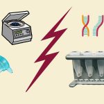
Several methods can be used to separate exosome from a body fluid (Figure 2), while most of the exosome separation methods are based on the ultra-centrifugation (UC) technique (Brennan et al., 2020). Some of the exosome isolation methods are summarized here:
Ultracentrifugation based isolation: Exosomes can be separated through series of centrifugation technique, by removing unnecessary components such as cells and cell debris. These centrifugations will provide a supernatant of exosomes. Finally, ultracentrifugation will provide a pellet of exosomes. But it is very important to wash pelleted exosomes with cell culture media or PBS at least once to purify them. It is also important to perform all centrifugation steps at 4°C in order to keep proteases, DNases and RNases inactive.
Ultracentrifugation with sucrose density gradients: For this technique, the exosome pellet will further be centrifuged at 100,000–200,000 x G for 120 min in a sucrose gradient medium. Exosomes should float at sucrose densities ranging from 1.15 to 1.19 g/mL on continuous sucrose gradients. Although this method provides a highly purified exosome, it is time-consuming and labor intensive. Also, the setup of centrifuging speed and time requires highly experienced personals.

Figure 2: Schematic summary of exosome isolation using five different methods from human serum and downstream analyses. With Western blot (WB) or nanoparticle tracking analysis (NTA) (Brennan et al., 2020).
Cushion ultracentrifugation (CUC): The serum sample needs to be diluted in 4.6 ml PBS and 0.4 ml of 40% iodixanol-PBS for ultracentrifugation at 120000 g for 2 hours at 4 °C. After centrifugations, the exosome pellet requires resuspension in 50–100 µl residual PBS for CUC.
Size-exclusion chromatography (SEC) based isolation: It is an ultrafiltration method that is faster than ultracentrifugation although exosomes might aggregate and clog the gel pores resulting in low yield. Also, non-exosomal RNA contamination is also an issue for using this technique. Therefore, it requires a washout step after each filtration step to improve exosome recovery. A nano-filtration concentrator might be useful for rapid exosome enrichment with short periods of centrifugation (Ludwig, Razzo, Yerneni, & Whiteside, 2019).
Precipitation based isolation: Exosomes can easily precipitate in several volume-excluding polymer solutions, for example, polyethylene glycol. If the sample incubates overnight with polyethylene glycol at 4°C, then low-speed centrifugation provides an exosome pellet. Also, lectins could be useful by agglutinate exosomes and pellet them at relatively low centrifugal speed (Chang, Chang, Chao, & Yu, 2018).
Immuno-affinity-based isolation: Exosomes contain a high number of proteins including cell receptors on their surfaces. This allows them to bind with antibodies specific to their aptamers. Antibodies specific to exosomal protein markers, such as Hsp70, CD9, CD63, CD81, CD82, Rab-5b and TSG101 are used for this technique to select the desired exosome or to trap unwanted exosome. After entrapping the exosome with a specific anti-marker antibody, these antibodies can immobilize on ELISA plates, magnetic and chromatography beads (Cai et al., 2018), or use them in microfluidic devices.
Utilizing the immuno-partiality based on magnetic beads, these subsets can be specifically caught and examined using flow cytometry or potentially electron microscopy. For exosomes to go about as a marker, the vesicles should be enhanced and specially fractionated. Enhancement of exosomes largely done by ultracentrifugation and regularly in mix with sucrose thickness inclination. It separates subpopulations of exosomes if present. The proteins CD9 and CD81 are normal extracellular marker which can be utilized for specific catch. Latex beads for specific capture require multiple centrifugation steps which poses a challenge. Nano-sized magnetic beads are an alternative to latex beads for exosome catch, detaching a more homogenous populace of vesicles concerning size and protein content. These beads require ultracentrifugation to reduce sample volume. Flow cytometry could not be done as they are too small to be detected from this process. Exosome isolation efficiency is influenced by factors such as incubation time, temperature, level of surface marker expression, and concentration of target vesicles.
The nature and state of the target molecule/structure, characteristics of the antibody–antigen interaction, sample type, concentration and ratio of beads and target molecules have an influence on the success of the magnetic separation. Magnetic beads coated with antibodies specific for exosome surface proteins in the isolation protocol after pre-enrichment further increases the purity of the exosome preparation. Direct exosome isolation with magnetic beads excludes the pre-enrichment step. Direct immunomagnetic isolation produces highly purified exosome preparations with minimal loss and requires less time. Direct isolation depends on the content of exosomes in the starting material and is determined by flow cytometry. If the signal is very low, indicating a very low exosome concentration in the sample a pre-enrichment step is needed. Low pH and high salt have been used to release protein complexes from the beads. The exosomes can be processed in the presence of the beads if the downstream application is mass spectrometry.