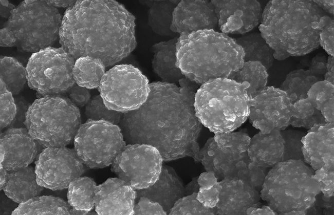
magnetofection, also known as magnetic transfection, is a procedure in which nucleic acid is bound to magnetic nanoparticles (MNPs) followed by their delivery into eukaryotic cells through applying magnetic force. The MNPs are usually made up of iron oxide and degraded within living cells, thus posing no hazard to cells. This technique has broader applications in molecular, cellular biology research and potential therapeutic applications (1).
How Magnetofection Works
In Magnetofection, nucleic acids are first bound to MNPs, followed by their delivery into eukaryotic cells with the help of a magnetic field. The binding of nucleic acids [deoxyribonucleic acid (DNA) and ribonucleic acid (RNA)] to MNPs is facilitated through electrostatic interactions. Both the DNA and RNA have negative surface charges and are attracted towards positive (cationic) materials on the surface of MNPs. The in vitro and in vivo experiments proved that nucleic acid molecules bound to MNPs are transferred inside the cell through magnetofection in a highly efficient manner (2).
Magnetofection Advantages Over Other Transfection Methods
Transfection is a procedure for delivering nucleic acids inside the eukaryotic cells utilizing either physical or chemical methods. Viral vectors mediated nucleic acid transfer into the cells is classified as transduction; however, sometimes their delivery is mediated through transfection methodologies. Magnetofection, including several other methods like microinjection, sonoporation, electroporation, gene gun, nucleofection, microneedles, and magnetoporation, fall in the category of physical methods. Chemical transfection methods are mainly lipid or polymers-based. Several modifications of chemical methodologies like nanoparticles, quantum dots, and graphene-mediated nucleic acid delivery with silica nanoparticles or carbon nanotubes are also used in some protocols. However, among all these, magnetofection is relatively more straightforward in use, efficient in delivering nucleic acid, and economical compared with all other methodologies that involve sophisticated equipment and complex formulations.
Delivery of free nucleic acids or therapeutic gene containing vectors into the eukaryotic cells/tissues is quite challenging. In intracellular environments, degradative enzymes impede delivering therapeutic genetic materials inside the cells/tissues. Magnetofection has been recognized as one of the most powerful tools for delivering intact/functional nucleic acids and vectors containing specific genetic material being used, thus having the potentials for correcting dysfunctional gene (3).
Suppose magnetofection is being utilized for the delivery of RNA. In that case, it should get transferred into the cellular cytoplasm for expression. In contrast, DNA first goes into the nucleus, followed by forming messenger RNA that goes out to the cytoplasm for expressing the desired product.
Several scientific reports have provided proof of concept about the superiority of magnetofection compared with other transfection procedures. According to a study in vitro gene transfer into the microglial cells that protect our brain, magnetofection efficiently transferred genetic material compared with conventional gene transfer methodologies (4).
Recent Advancements in Magnetofection:
Magnetofection has great application in gene therapy protocols in which defective genes are replaced with healthy ones into the human cell/tissues. Compared with other protocols, magnetofection mediated delivery of healthy genes into human cells is considered most promising. However, there is ongoing research to develop a proper formulation and appropriate magnetic field skills. Gene therapy through magnetofection for therapeutic purposes besides targeted delivery offers the unique advantage of being least invasive (5).
Magnetic nanoparticles mediated nucleic acid transfer into stem cells (6), and the flexibility of methodology (7) add to its versatility for its extensive utilization in regenerative medicine and genetic diseases.
Magnetofection in Clinical Practice
It has become a fact that magnetofection if properly used, could efficiently transfer nucleic acids as well as therapeutic genes containing viral vectors into the human cells/tissues. However, its development for clinical usage is hampered due to the chemical nature of MNPs fate in vivo. Research is ongoing on the bioevaluation of MNPs for their potential clinical use in gene therapy protocols involving magnetofection (8). As far as clinical applications of magnetofection are concerned, targeted delivery into cancerous tissues for controlling several cancer types holds a tremendous future promise in clinics (9).
Sources:
- Plank C, Anton M, Rudolph C, Rosenecker J, Krotz F. Enhancing and targeting nucleic acid delivery by magnetic force. Expert Opin Biol Ther. 2003;3(5):745-58.
- Bi Q, Song X, Hu A, Luo T, Jin R, Ai H, et al. Magnetofection: Magic magnetic nanoparticles for efficient gene delivery. Chinese Chemical Letters – doi: https://doiorg/101016/jcclet202007030. 2020.
- Scherer F, Anton M, Schillinger U, Henke J, Bergemann C, Kruger A, et al. Magnetofection: enhancing and targeting gene delivery by magnetic force in vitro and in vivo. Gene Ther. 2002;9(2):102-9.
- Smolders S, Kessels S, Smolders SM, Poulhes F, Zelphati O, Sapet C, et al. Magnetofection is superior to other chemical transfection methods in a microglial cell line. J Neurosci Methods. 2018;293:169-73.
- Qunjie B, Song X, Hu A, Luo T, Jin R, Ai H, et al. Magnetofection: Magic magnetic nanoparticles for efficient gene delivery[J]. . Chinese Chemical Letters. 2020;31(12):3041-6.
- Yamoah MA, Thai PN, Zhang X. Transgene Delivery to Human Induced Pluripotent Stem Cells Using Nanoparticles. Pharmaceuticals. 2021;14(4):334.
- Blokpoel Ferreras LA, Chan SY, Vazquez Reina S, Dixon JE. Rapidly Transducing and Spatially Localized Magnetofection Using Peptide-Mediated Non-Viral Gene Delivery Based on Iron Oxide Nanoparticles. ACS Appl Nano Mater. 2021;4(1):167-81.
- Dragar C, Kralj S, Kocbek P. Bioevaluation methods for iron-oxide-based magnetic nanoparticles. Int J Pharm. 2021;597:120348.
- Belete TM. The Current Status of Gene Therapy for the Treatment of Cancer. Biologics. 2021;15:67-77.